


Gallery
Finger Ganglion – Mucous cyst

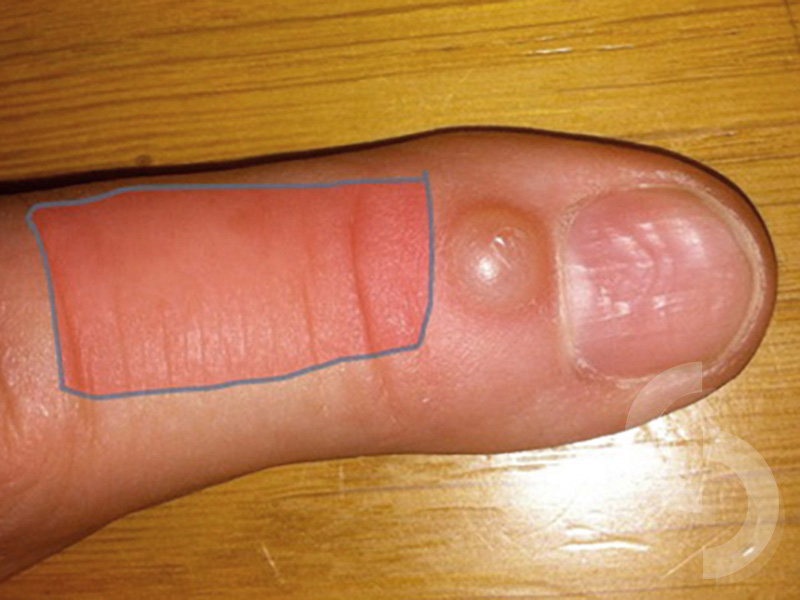
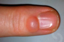
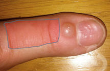
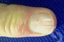
Picture 1 - characteristic nail grooving due to compression of the nail root with the ganglion
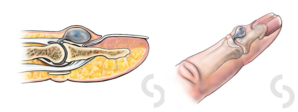
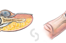
Picture 2 - Anatomy of the fingertip and common ganglion location - majority of these ganglions originate from the joint
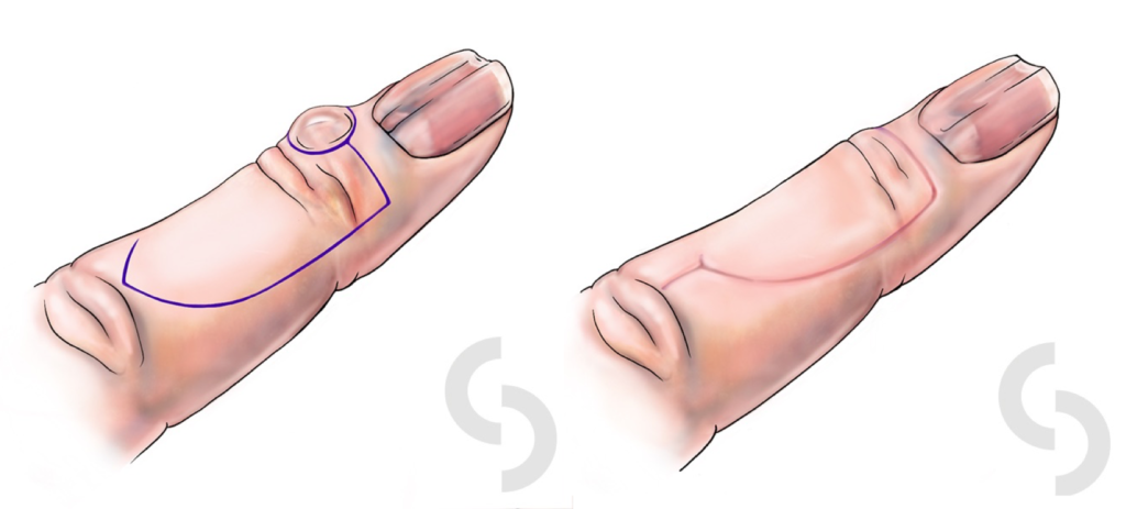
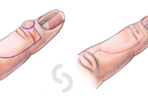
Picture 3a - Skin flap often has to be mobilised from the nearby finger area in order to cover the defect created by adequate cyst excision
Picture 3b - Area of the finger from which extra skin is 'borrowed' to compensate for skin shortage
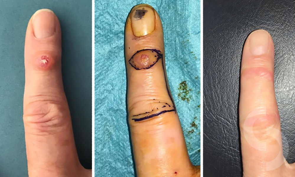
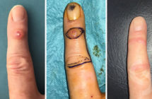
Picture 4 - LEFT - ganglion which involves the overlying skin; MIDDLE - marking of the ganglion and the skin flap before the operation; RIGHT - 3 weeks after surgery
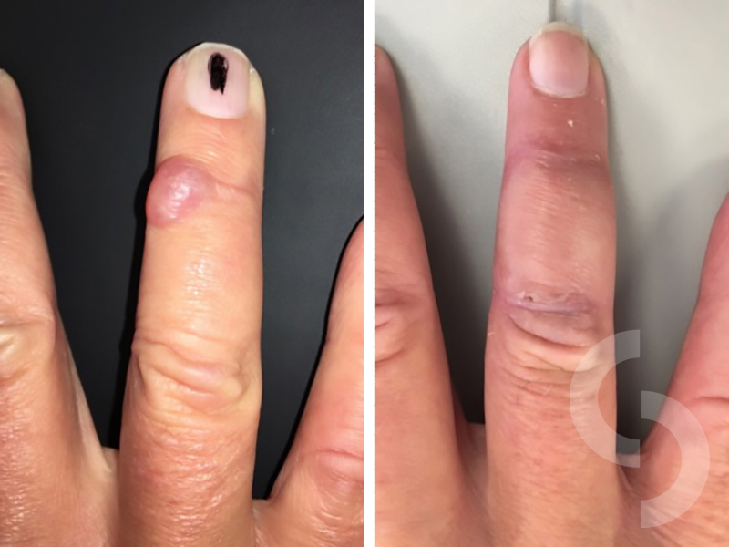
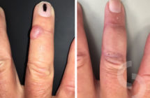
Picture 5 - Before and 3 weeks after ganglion removal and reconstruction with a skin flap

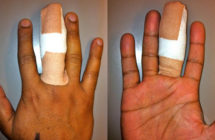
Picture 6 - Finger dressings to be worn over the first 2 weeks after surgery

Picture 7 - Improvement in the nail surface 2 months after surgery i.e. 'decompression' of the nail root by ganglion removal
I am grateful to all patients who have kindly agreed for their pictures to be used here.