


Gallery
Kienbock’s Disease

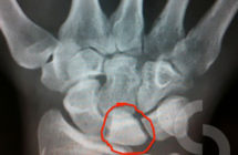
Position of the lunate bone within the wrist joint; This X-rays image shows stage II Kienbock's disease
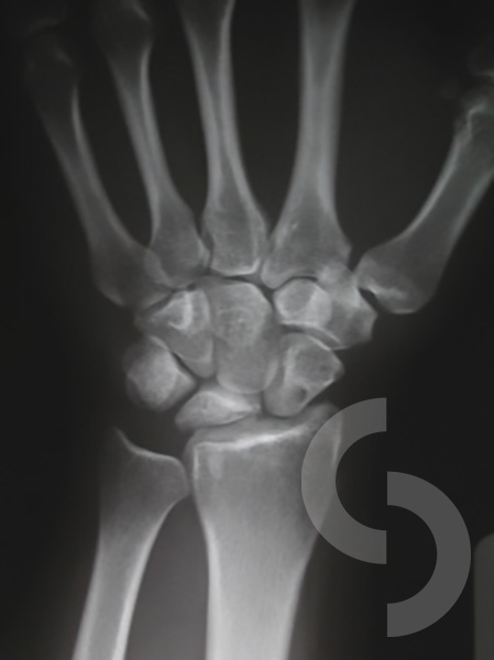
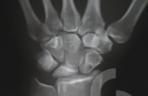
Stage III Kienbock's disease - further 'flattening' of the bone

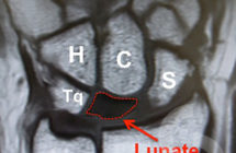
MRI of the wrist showing abnormal lunate (reduced height of the bone) and its position against neighbouring wrist bones (Scaphoid, Capitate, Hamate and Triquetrum) and forearm bones (Radius and Ulna)
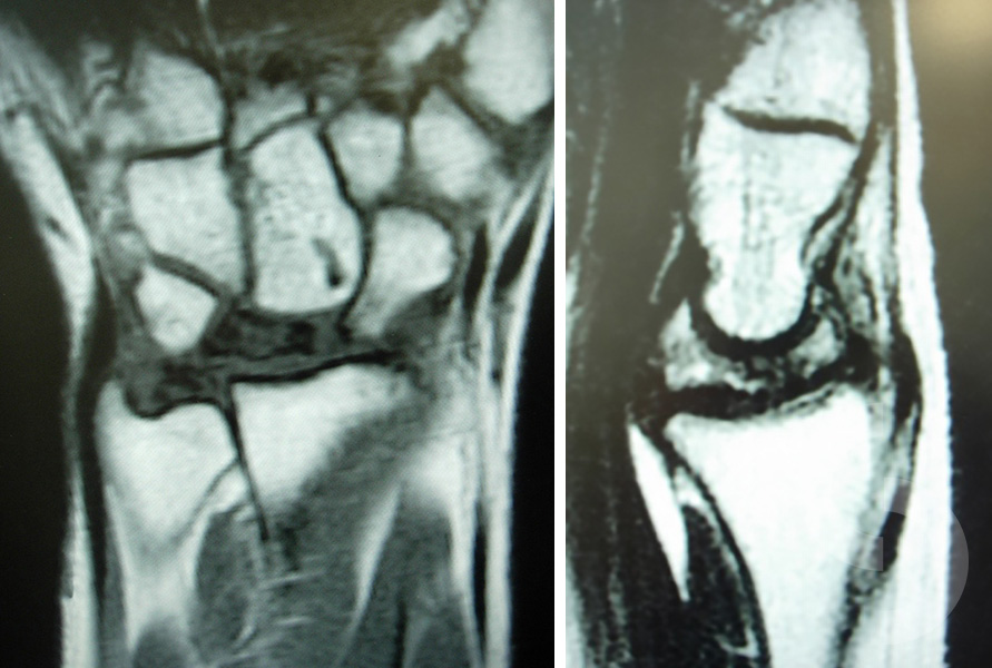
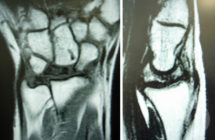
MRI - Lunate collapse/fragmentation evident in advanced stages of Kienbock's disease
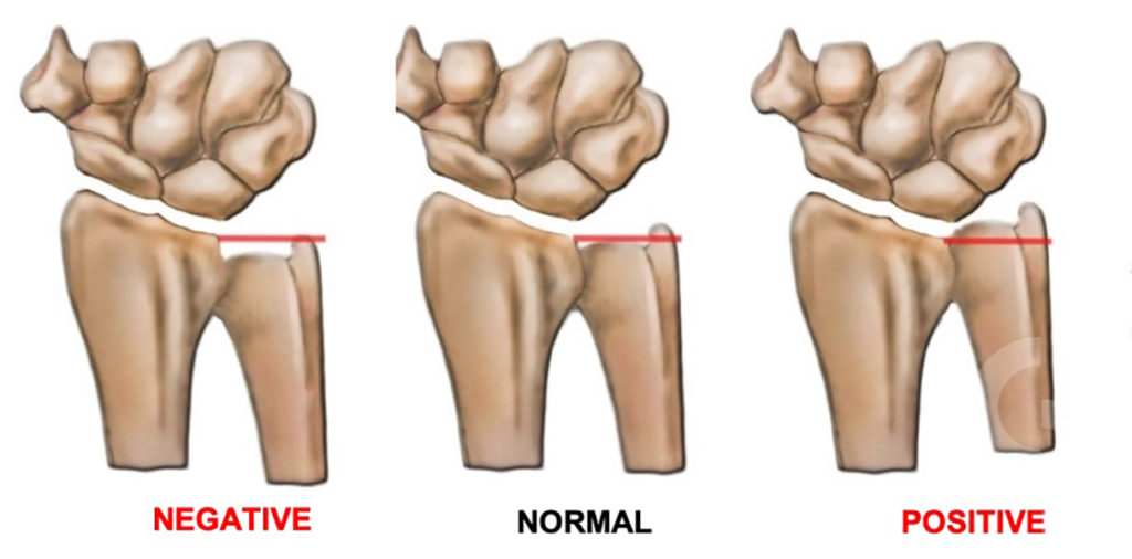
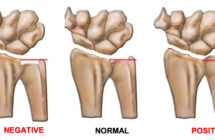
Ulna Variance = relative lengths of the joint surfaces of the radius and ulna. NORMAL or NEUTRAL - both bones joint surfaces are at the same level; NEGATIVE - ulna 'appears short', projects less distally; POSITIVE - ulna 'appears longer', projects more distally
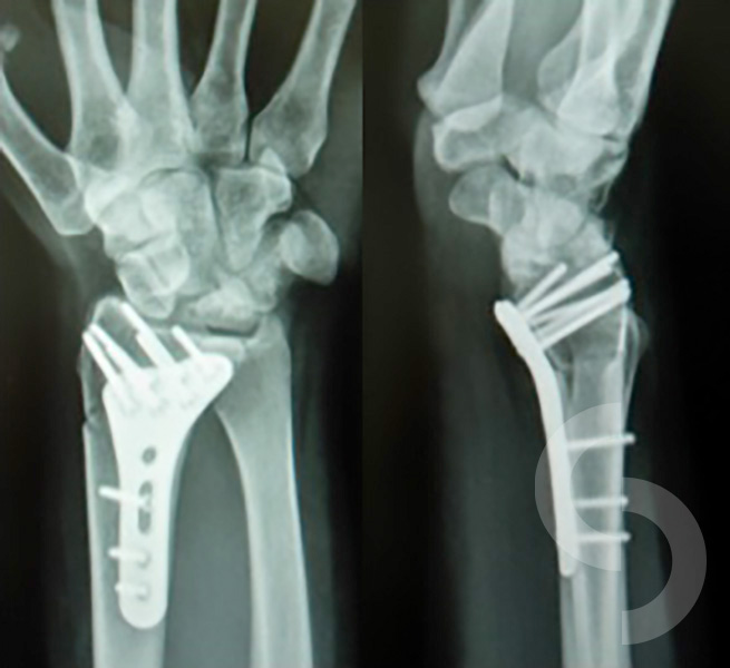
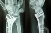
After radial shortening performed to level the radius joint surface more favourably against 'shorter' ulna (common treatment for the negative ulna variance)
I am grateful to all patients who have kindly agreed for their pictures to be used here.