


Gallery
Scaphoid Fractures and Non-unions

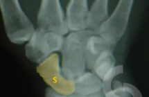
Wrist X-rays - position of the scaphoid (S) bone within the wrist joint
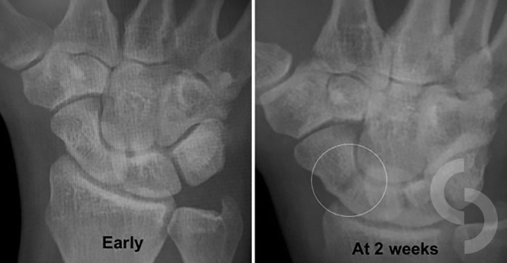
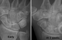
Scaphoid fractures are often not obvious early and only become visible later, usually on repeated X-rays 1 -2 weeks later
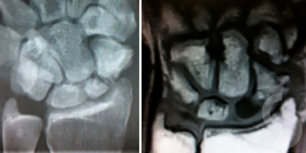
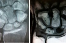
Scaphoid fracture can be associated with unfavourable loss of vascularity (avascular necrosis) to one of its fragments; MRI is required to delineate this as demonstrated on the right picure: marked colour discrepancy between two fragments; black part of broken scaphoid on this image confirms avascular necrosis
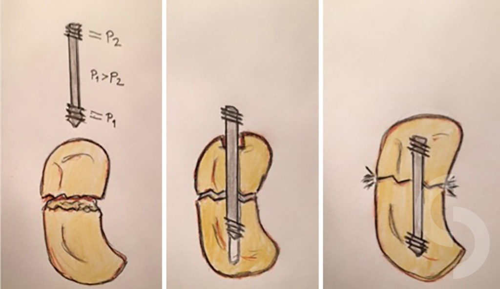
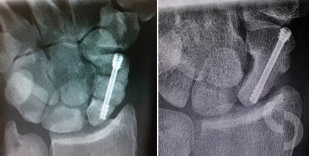
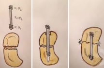
Special compression screw is used in surgical fixation of two scaphoid fragments to improve chances of their healing (union)

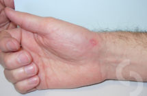
Tiny scar 3 weeks after the percutaneous fixation of the scaphoid fracture with compression screw
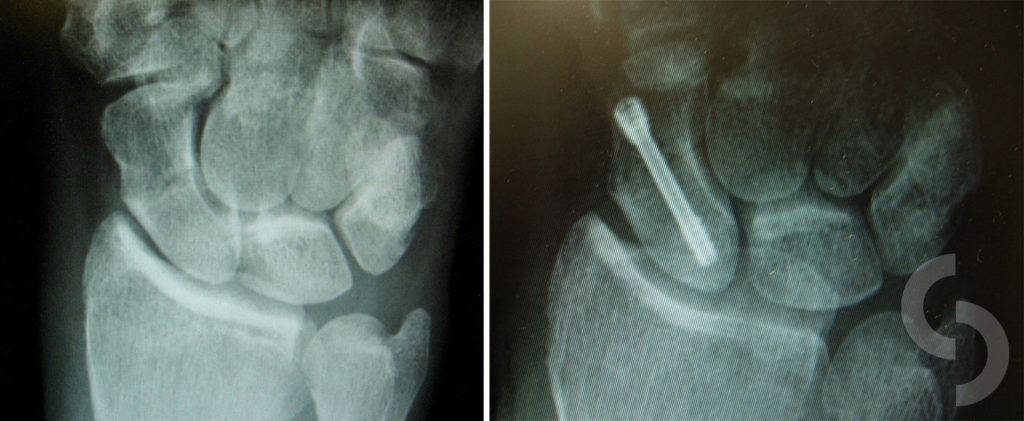
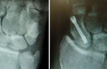
Example of good scaphoid union with compression screw fixation
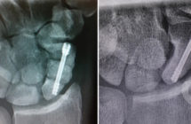
Scaphoid non-union treated with the bone graft inserted between two non-healing scaphoid fragments; all three fixed with compression screw
I am grateful to all patients who have kindly agreed for their pictures to be used here.