Gallery
Wrist Ligament Injuries
![](https://sonjacerovac.com/wp-content/uploads/2020/04/Wrist-ligaments-watermarked-1024x611.png)
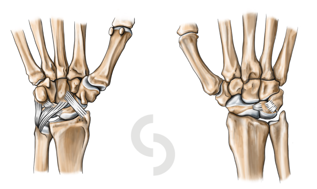
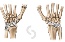
Picture 1: Schematic representation of complex anatomical arrangement of few extrenal and internal wrist ligaments
![](https://sonjacerovac.com/wp-content/uploads/2019/02/SLL-tear-1024x417.jpg)
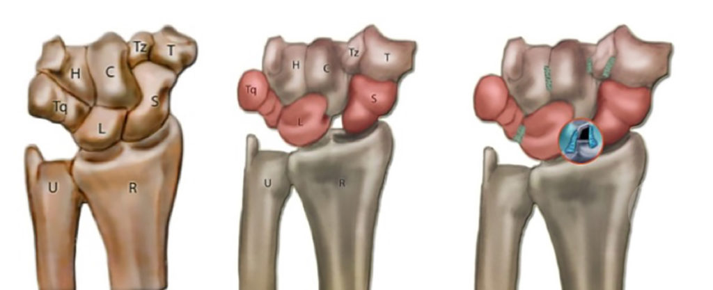
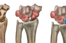
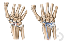
Picture 2: Left - normal alignment of 8 wrist bones; Right: disruption of the ligament between the scaphoid (S) and lunate (L) and consequent gap between them
![](https://sonjacerovac.com/wp-content/uploads/2019/02/Hug.jpg)
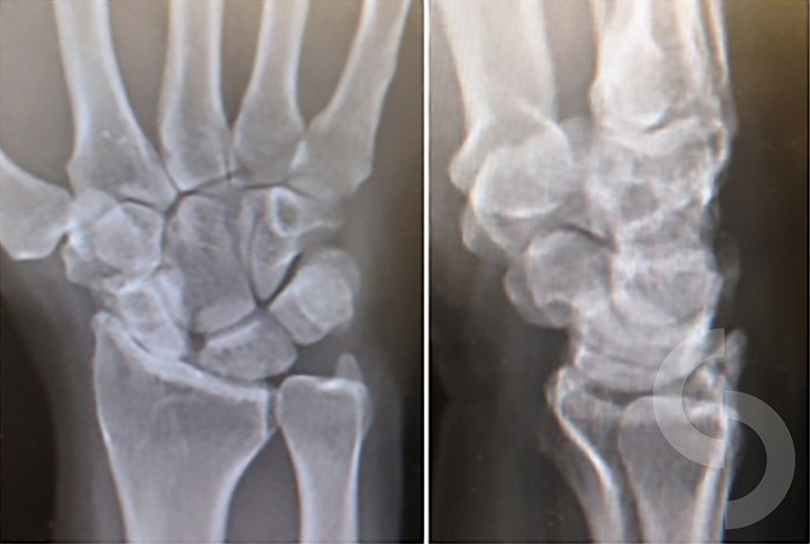
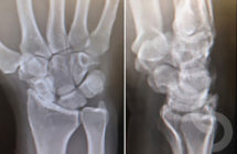
Picture 3: X-rays image of advanced arthritis caused by malrotation of the wrist bones after crucial ligament rupture
![](https://sonjacerovac.com/wp-content/uploads/2019/02/MRI-SLAC.jpg)
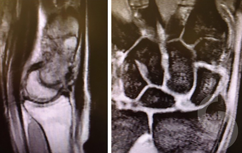
Picture 4: MRI image indicating increased gap between scaphoid and lunate after the tear of the ligament between them
![](https://sonjacerovac.com/wp-content/uploads/2020/01/SLL-and-Brunelli-2-1024x686.png)
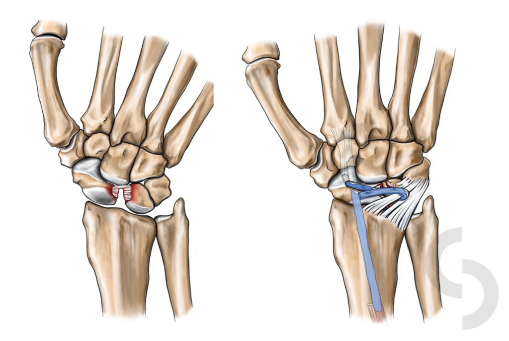
Picture 5: Reconstruction of torn scapho-lunate ligament with a flexor carpi radialis tendon sling taken from the forearm and transferred into the wrist
![](https://sonjacerovac.com/wp-content/uploads/2020/04/Wrist-ligaments-watermarked-215x140.png)
![](https://sonjacerovac.com/wp-content/uploads/2019/02/SLL-tear-215x140.jpg)
![](https://sonjacerovac.com/wp-content/uploads/2019/02/Hug-215x140.jpg)
![](https://sonjacerovac.com/wp-content/uploads/2019/02/MRI-SLAC-215x140.jpg)
![](https://sonjacerovac.com/wp-content/uploads/2020/01/SLL-and-Brunelli-2-215x140.png)
I am grateful to all patients who have kindly agreed for their pictures to be used here.